|
| |
|
|
|
|
|
|
|
|
|
|
|
|
|
|
|
|
|
|
|
|
|
| |
 |
| |
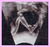
scan of a foetus
|
| |
|
| |
|
| |
|
| |
|
| |
|
| |
|
| |
|
| |
|
| |
|
| |
|
| |
|
| |
|
| |
|
| |

|
| |
scan of the baby's
face
|
| |
|
| |
|
| |
|
| |
|
| |
|
| |
|
| |
|
| |
|
| |
|
| |
|
| |
|
| |
|
| |
|
| |
|
| |
|
| |
scan of the baby's leg
|
|
|
|
|
|
|
|
|
|
|
|
|
|
|
|
|
|
|
|
|
|
|
|
|
|
|
|
|
|
|
|
|
|
|
|
|
|
|
|
|
|
PRENATAL
CHECKUPS
|
|
|
|
|
 |
THE
FIRST COMPULSORY VISIT : |
|
|
|
It has to be
done before week 14, that is to say before the third month of
pregnancy. However the earlier you do it, the best it is. In fact as
soon as you know or suspect to be pregnant go and see your doctor or
gynaecologist. Indeed it is better to know the results of the urine
and blood tests asap, in case of problems.
The first visit will be a complete one
: you will have to answer questions
about you and your family medical background, but also this of your
partner. It will be followed by a physical examination and then
prescription of your analyses .
|
|
|
|
THE DISCUSSION : |
|
|
|

|
your medical background :
you will be asked questions about your age, your childhood illnesses,
the surgical operations you underwent, if you suffer from allergies,
if you take medicines....
|
|
|
|
|
your family medical history and this
of your partner : if there is a
recurrent illness in your family, like diabetes or hypertension. Also
if there are twins in your family or your partner's one (it can
hereditary).
|
|
|
|
|
your gynaecology background :
questions will be about your LMP date (last
menstrual period), your cycle, about your other pregnancy if any,
about miscarriage or abortion if any. |
|
|
|
|
|
|
THE PHYSICAL
EXAMINATION : |
|
|
|
Some of you may be surprised that there is no nurse with the
doctor when he examines you. You generally undress in front of him, and no gown will be given
to you. |
|
|
|
|
|
you will be weighed. Indeed
weight control will be strictly followed by doctors during your
pregnancy. Rapid loss or gain weight will be investigated. A loss of
weight during the first trimester can be explain by frequent nausea
and vomiting (see morning sickness). For gain weight it can be due
because you eat too much ( reminder : you don't need to eat for two)
but it can also be due to your thyroid gland.
|
|
|
|
|
your blood pressure will be measured.
Normal one is 12/7. Control will be done
throughout your pregnancy to detect hypertension.
|
|
|
|
|
your general physical health will be
checked. Your heart and lung will be
listened to, your teeth looked at,... |
|
|
|
|
|
|
The
blood and urine prescription : |
|
|
|
|
|
the laboratory urine test : to
check the sugar and albumin levels in your blood (French term :
Glycosurie, Protéinurie / Albumine).
|
|
|
|
|
the blood test : researched
elements are blood and RH group, rubella (German measles), syphilis,
Aids, toxoplasmosis, HCG level, hepatitis.
|
|
|
|
|
|
|
|
|
THE
SECOND VISIT : |
|
|
|
The
2nd prenatal compulsory visit takes place during your fourth
month of pregnancy. Since now, you will have one per month.
|
|
|
|
At
each visit some routine examinations will be done : |
|
|
|
|
|
checking of your blood pressure, to detect hypertension.
|
|
|
|
|
checking of your weight. You will be reminded at each visit that the
average weight gain for all the pregnancy is around 9-12kg.
|
|
|
|
|
checking of the height of your womb. Just to confirm that your uterus
goes on growing and going up towards your stomach. To measure
the height the doctor will put a centimetre on your abdomen, from the
pubic bone to the end of your uterus, called fundus. |
|
|
|
|
|

|
 At the 3rd month,the usually the height of the fundus is : 9 cm At the 3rd month,the usually the height of the fundus is : 9 cm
At the 5th month,the usually the height of the fundus is : 20 cm.
At
the 6th month,the usually the height of the fundus is : 24 cm.
At the 7th month,the usually the height of the fundus is : 28 cm
At the 8th month,the usually the height of the fundus is : 30 cm.
At the 9th month,the usually the height of the fundus is : 33 cm. |
| |
|
|
|
To make space it crowds out the
other organs, which leads again to heartburn at the end of the
pregnancy. At month 9 your uterus is against your breast bone which
emphasized your breathlessness. |
|
|
|
|
|
|
|
Special
examination to this 2nd visit of the 4th month :
|
|
|
|
|
|
The gynaecologist will speak to you of the risks of having an abnormal
baby and of tests to do to know it. At the first scan the radiologist
has already measured the nape of your baby's neck, which can give an
indication of abnormality.
Mongolism is the result of a
chromosomal abnormality : the foetus has an extra chromosome. The
cause of this abnormality is unknown. Doctors just know that the risk
is emphasized by the age of the mother, and by the heredity factor.
Analyses
are not compulsory : the doctor will explain mongolism to the parents,
and ask them if they agree to make the tests. The result comes around
3 or 4 weeks after, which is very long when you are waiting for the
final answer. Moreover, through the discussion with the gynaecologist, you will understand how much it is difficult to know
that the baby you are waiting for is abnormal. Therefore if you are
absolutely against abortion, even for medical reasons, don't ask for
the test.
|
|
|
|
|
There are two major tests which are done in France, even if the 1st
scan has revealed a normal size of the nape. However none of them can
be done without your authorisation. It is a blood test for searching
the presence of the hormone responsible for mongolism (it is called
HT21 analysis),and for searching alpha fetoprotein responsible for
brain development abnormality or for spina-bifida.
|
|
|
|
|
In case the risk of having a Down's syndrome baby is suspected either
by the 1st scan or by blood tests, an amniocentesis
will be prescribed to confirm the blood result.
|
|
|
|
|
|
|
|
|
THE
OTHER VISITS : |
|
|
|
The
following visits :
Each time you will go to see your gynaecologist
the same routine examinations will be done :
|
|
checking of your blood pressure, to detect hypertension.
|
|
|
|
|
checking of the height of your womb.
|
|
|
|
|
checking of your cervix. |
|
|
|
|
checking of your weight. |
|
|
|
|
checking of the baby's heart beats |
|
|
If your pregnancy is a normal one, there will
be no special exams.
|
|
|
|
|
|
ULTRASOUND
SCANS : |
|
|
|

|
|
An
ultrasound scan gives a picture of the foetus and of the placenta. You
can see on the screen of the doctor's computer your baby in utero. The
doctor will give you several pictures of your baby, sometimes the
pictures will even be in colour.
The scan takes about 10 minutes and is harmless. A jelly is put on
your abdomen and a transducer passed on it. |
| |
|
There are compulsory scans to do. There are paid back by the CPAM. If
your gynaecologist has a scan in his office, you will probably have a
scan nearly each time you will go to see him, and they may be
free. |
|
|
|
THE
FIRST SCAN :

|
It generally happens during the third month, around week 12. For this
one, you will be asked to drink 1 litre of water in order to have a
full bladder. This will not be necessary for the other scans.
|
|
|
|
|
It is not easy for a non-skilled eye to understand what it sees. That
's why dare asking the doctor to explain it to you.
|
|
|
The
first scan aims are :
|
|
to determine the age of the foetus (in case you do not know your LMP),
the measure of the head, and of the body.
|
|
|
|
|
to check how the foetus is growing and its vitality.
|
|
|
|
|
to measure the nape of the foetus' neck, which can be a hint about
mongolism. |
|
|
|
|
to see visible abnormality, such as no brain, one lung ... |
|
|
|
|
to find the position and the condition of the placenta |
|
|
|
|
to see if you are waiting for one baby or twins.
|
|
|
|
|
|
|
THE
SECOND SCAN :
|
|
It generally happens during the second trimester, between week 18
& 24. This scan is especially a morphologic one, which aim is to
take all the measures of the foetus. The most relevant measures are
these of :
|
|
|
|
|
the head diameter (called BIP = diamètre bipariétal = parietal
diameter)
|
|
|
|
|
the length of the femur
|
|
|
|
|
the length of the feet |
|
|
The practitioner also checks the presence and
the development of :
|
|
the lips
|
|
|
kidneys
|
|
|
liver |
|
|
stomach |
|
|
arms |
If you don't want to know if you are waiting for a boy or a girl tell
it to the doctor before he starts. It is during this second scan that
you can see it. |
|
|
|
THE
THIRD SCAN :
It generally happens during the third
trimester, during the eighth month. Its aims are :
|
|
to confirm that the cervix is closed.
|
|
|
|
|
to see if the baby is already head down. If she is not, don't worry
she can still have enough place to do it.
|
|
|
|
|
to see the baby's development |
|
|
|
|
to see the position of the placenta. Indeed in case of placenta
praevia, which means that the placenta is over or near the cervix, a
cesarean may be probably envisaged. |
|
|
|
|
the amount of amniotic fluid |
|
|
|
|
the beats of the baby's heart.
|
|
|
|
|
|
|
AMNIOCENTESIS : |
|
|
|
Why
having amniocentesis?
|
|
Amniocentesis is a test done for detecting
some genetic defects such as spina-bifida or Down's syndrome. It is
also be prescribed when the gynaecologist suspects a foetus
abnormality or when there is a case of it in the parents' family
history.
|
|
|
|
|
Women over 35 are proposed to have amniocentesis because the age of
the mother may be an increasing risk factor of having a baby with
genetic defects.
|
|
|
|
|
When the HT21 test is positive, the
gynaecologist proposes an amniocentesis to confirm the previous test. |
|
|
|
Risk
of amniocentesis :
|
|
The risk may be a miscarriage. Of course this
risk is taken into account and weighted by the gynaecologist when he
offers you an amniocentesis . If you don't belong to potential risky
people, your obstetrician won't offer it.
|
|
|
How
does it work ? :
|
|
Amniocentesis consists in extracting some
amniotic fluid from the womb. First of all, a scan is done to see the
exact position of the foetus and of the placenta. A local anaesthetic
is spread on your abdomen. Then the doctor inserts a long needle
surmounted with a syringe into your uterus. The extracted fluid is
send to be analysed. It can takes two weeks to get the results.
Amniocentesis is not physically painful.
|
|
|
|
|
Amniocentesis is carried around weeks 16-18.
|
|
|
|
|
Karin,
our August interviewee, narrates her amniocentesis. |
Best viewed in
1024*768
All contents © toulousebabynet Feb 2002
IExplorer 5 & Netscape 4.7
|
